Long bone anatomy, structure, parts, function and fracture types
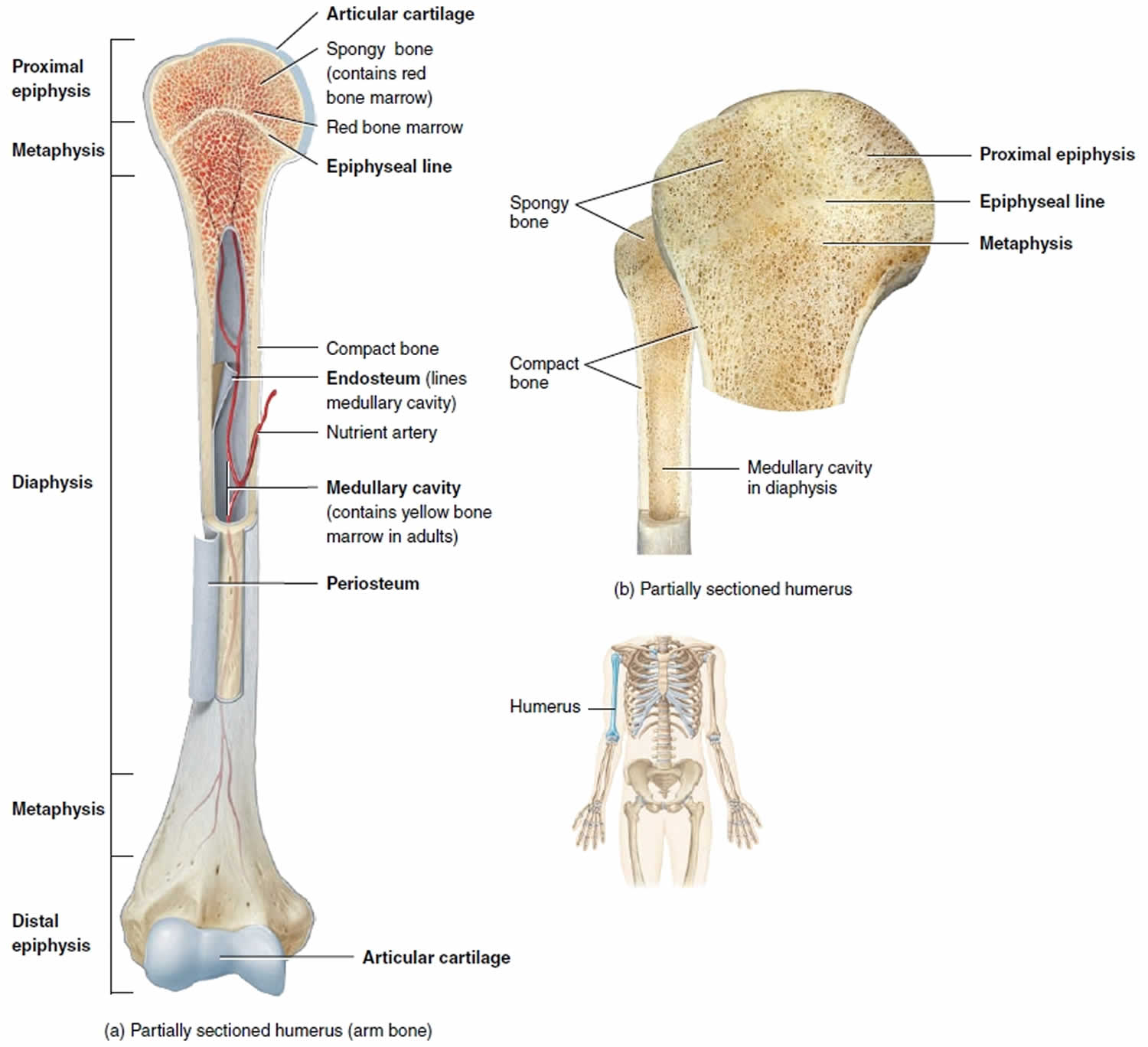
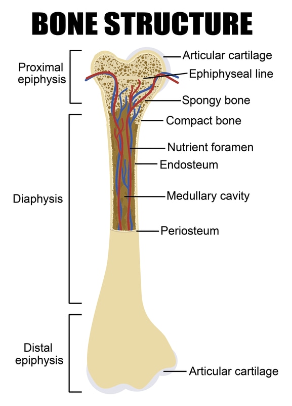
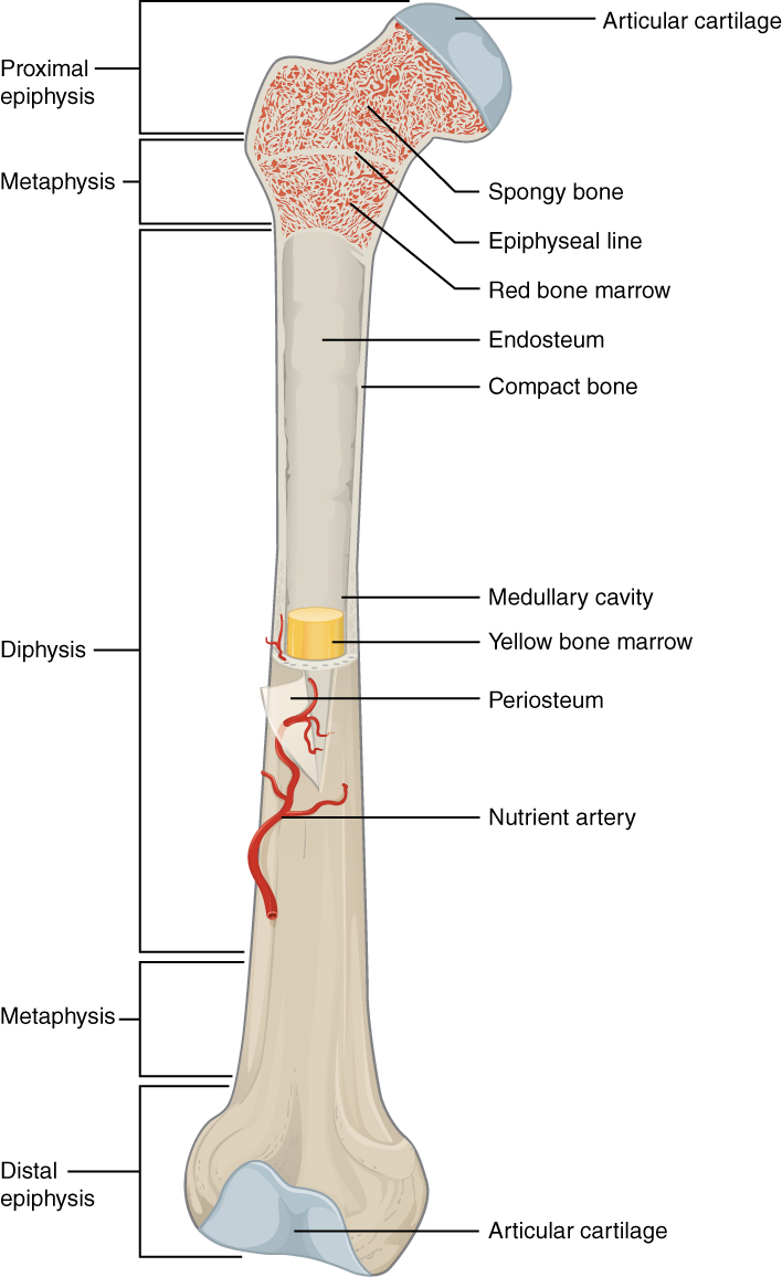
Figure 1. Anatomy of a Long Bone. A typical long bone shows the gross anatomical characteristics of bone. The structure of a long bone allows for the best visualization of all of the parts of a bone (Figure 1). A long bone has two parts: the diaphysis and the epiphysis.

parietal bone Colouring Pages (page 2) Anatomy bones, Skull anatomy
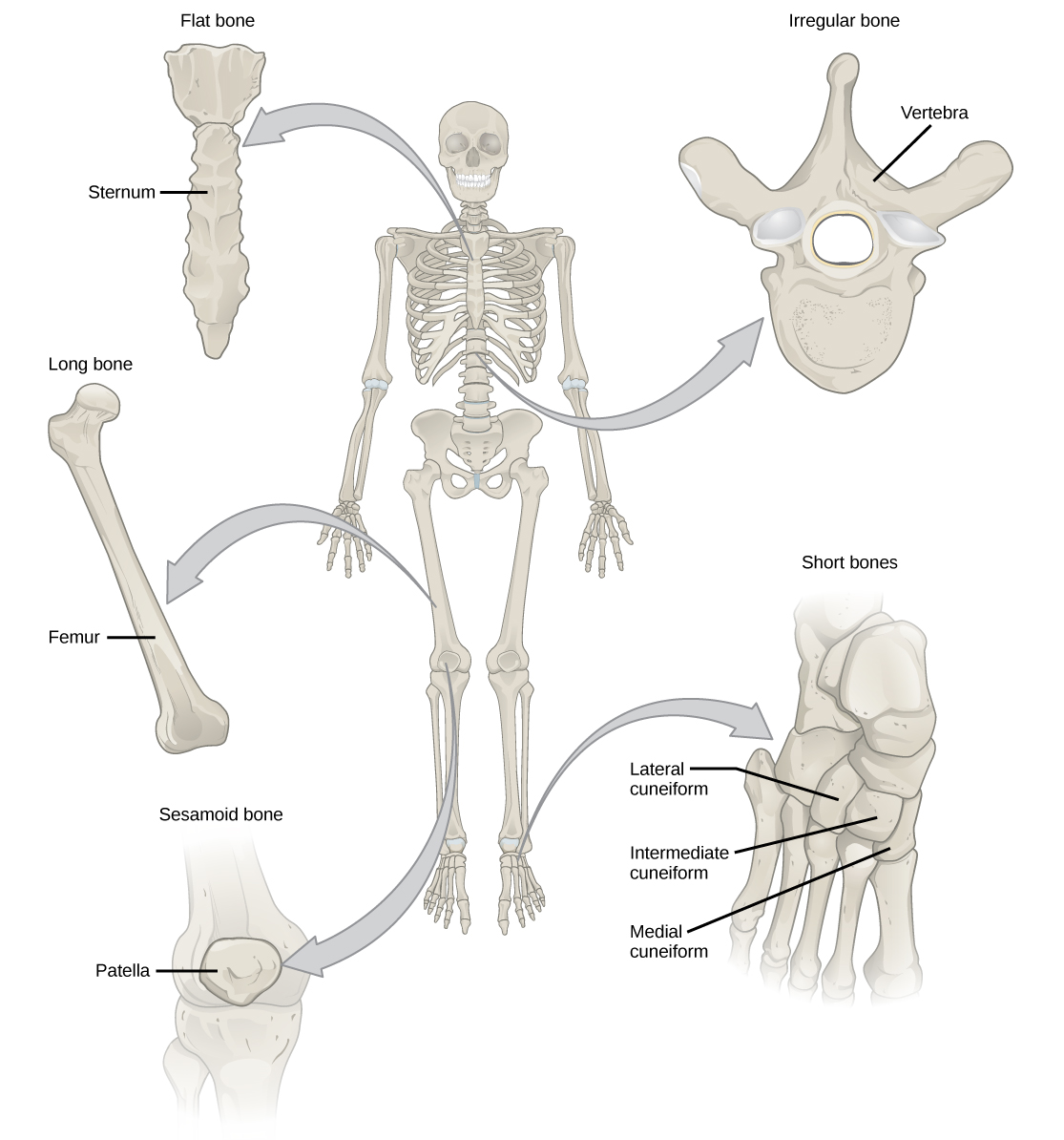
3. Label spongy bone structures shown in this micrograph (arrows): trabecula. bone marrow. 4. Identify the shape of the bones shown below as: long, short, flat, sesamoid or irregular. Write your answers on the spaces provided. 5. Name five bones of the axial skeleton and five bones of the appendicular skeleton.
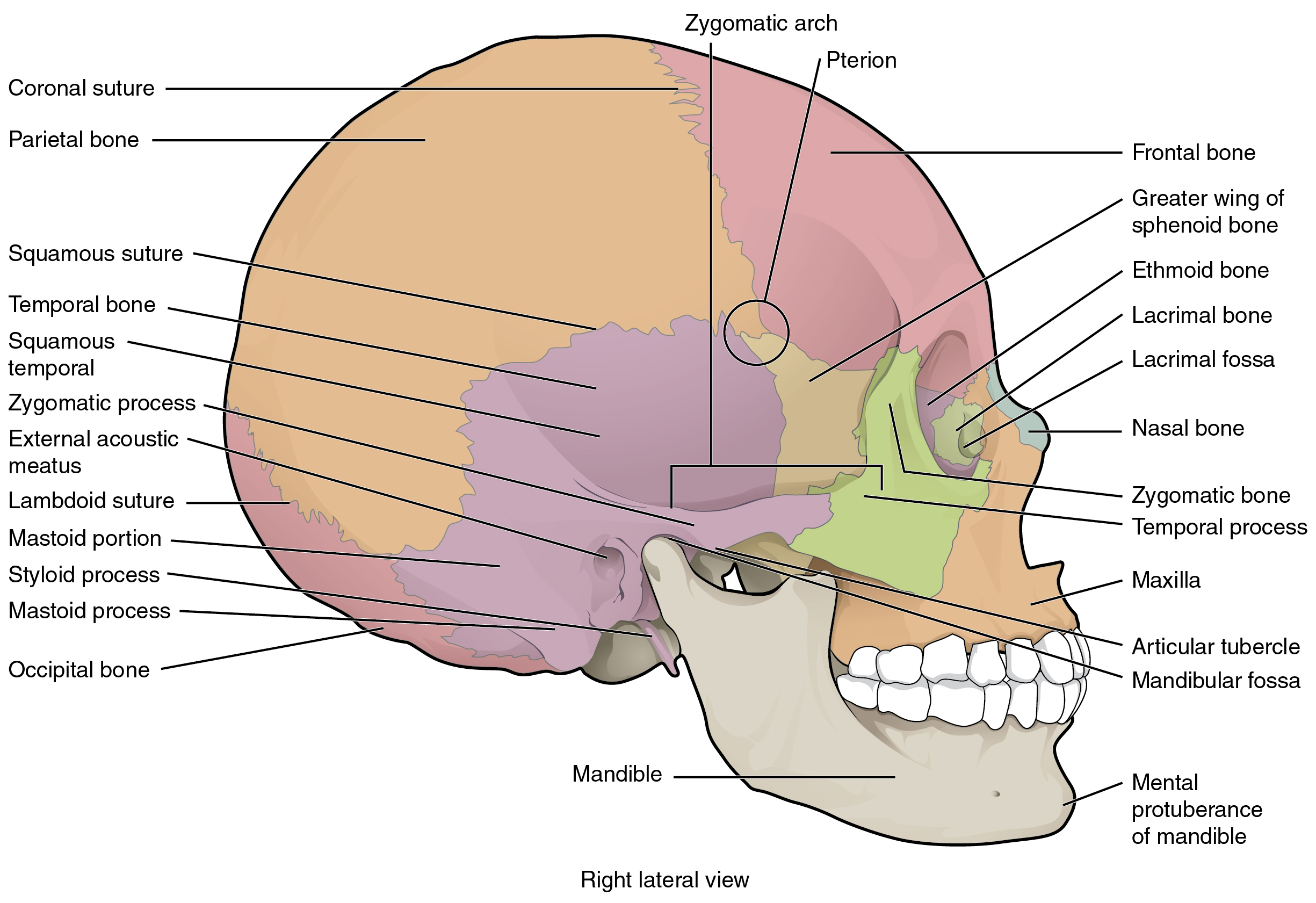
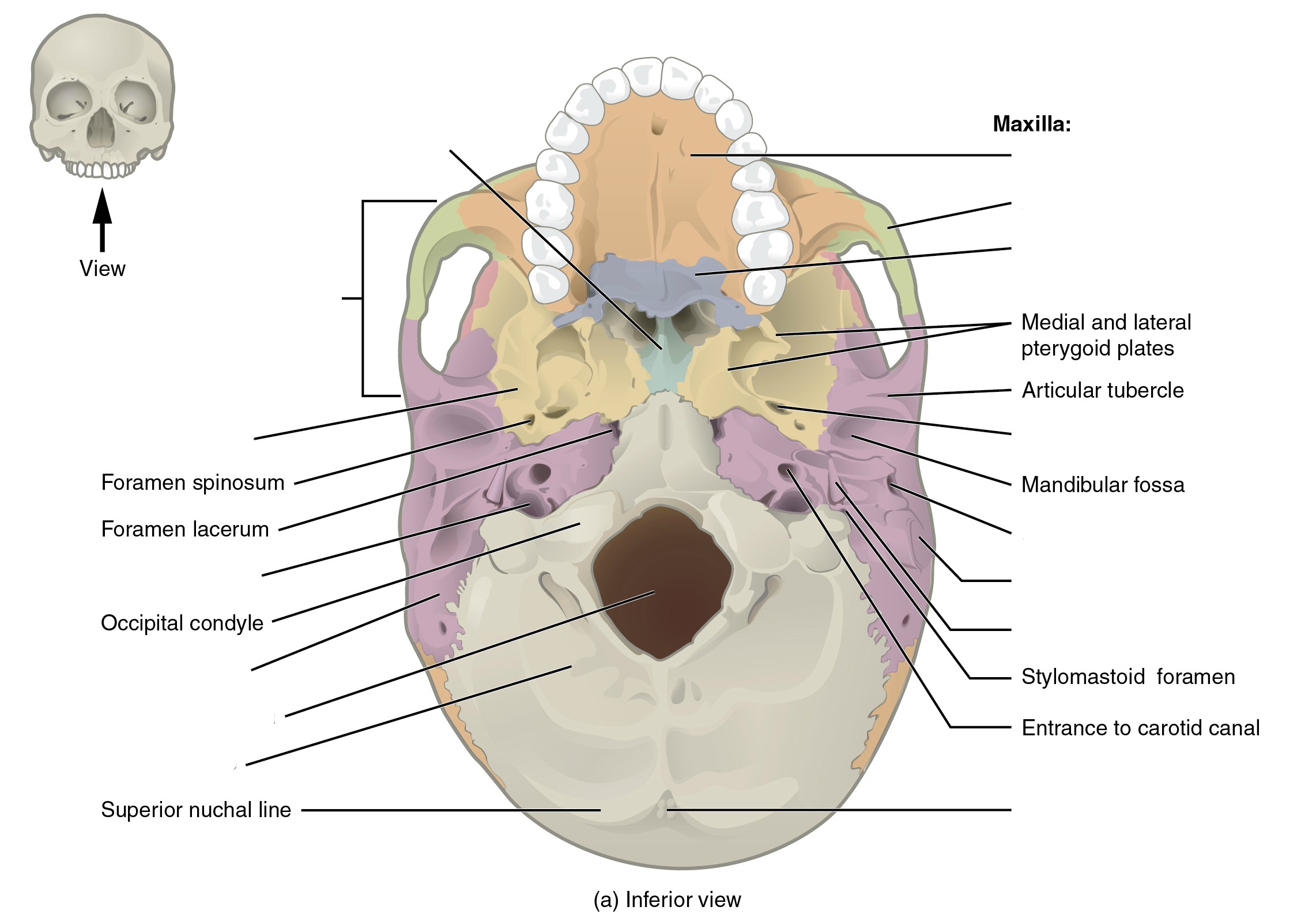
The Skull Anatomical Basis of Injury
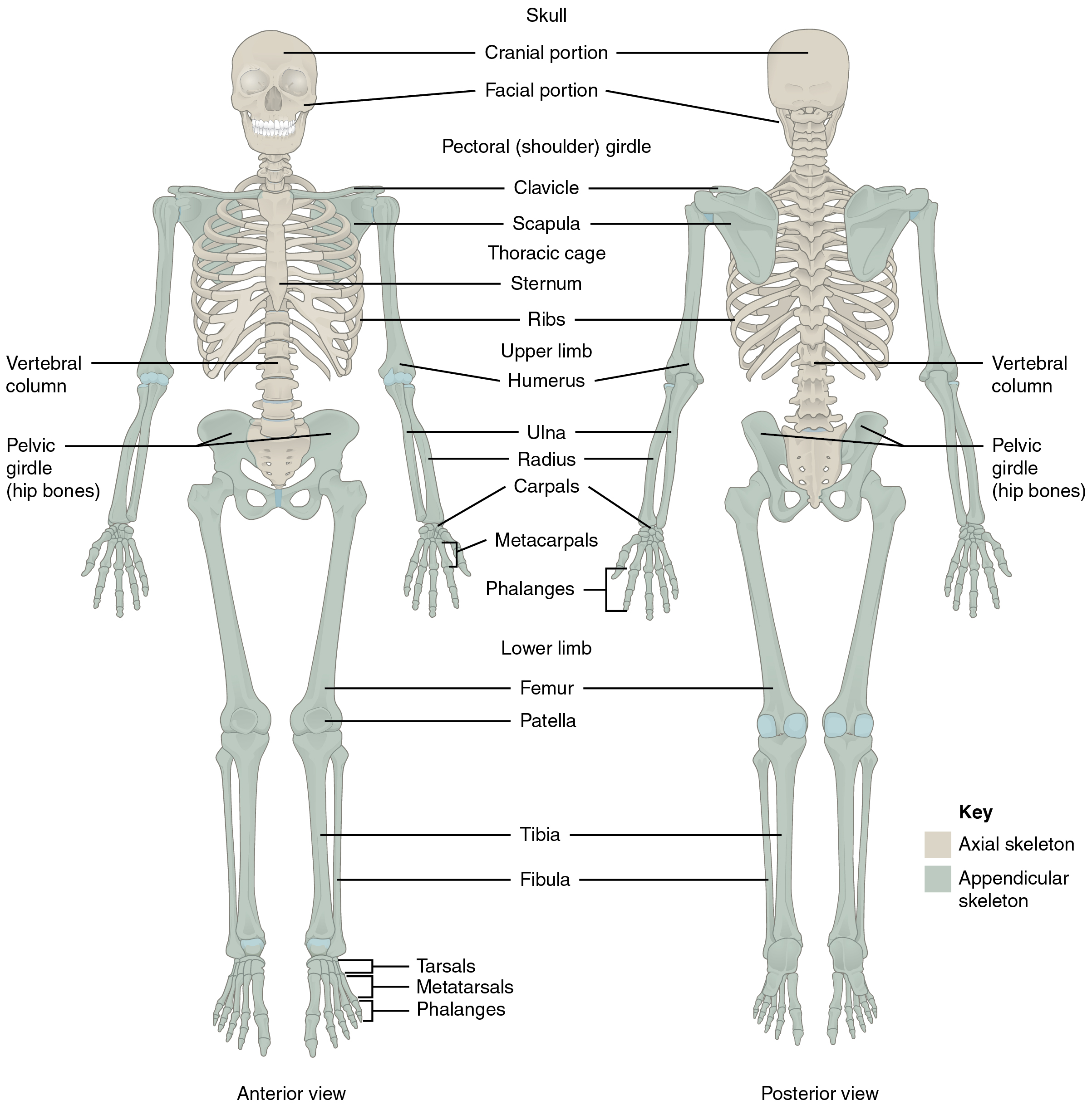
The human skeletal system consists of all of the bones, cartilage, tendons, and ligaments in the body. Altogether, the skeleton makes up about 20 percent of a person's body weight. An adult's.
Bone · Biology
The diaphysis is the hollow, tubular shaft that runs between the proximal and distal ends of the bone. Inside the diaphysis is the medullary cavity, which is filled with yellow bone marrow in an adult. The outer walls of the diaphysis (cortex, cortical bone) are composed of dense and hard compact bone, a form of osseous tissue.

Bones at California State University Sacramento StudyBlue
Differentiate between bones of the body based on the classification of the shape of the bone. 4. Identify the bones of the body using correct anatomical terminology. 5. Use correct anatomical terminology to correctly identify bone landmarks that serve as attachment points for skeletal muscles and ligaments. 6.

Skeletal System Anatomy and Physiology Skeletal system anatomy, Human
label the specific bony features of the skull in lateral view: label the bony structures of the shoulder and upper limb: label the bony structures of the thoracic cage: label the structures of the bone: Study with Quizlet and memorize flashcards containing terms like label the structures of a typical cervical vertebra:, label the structures of.

Pin by Karen Davies on anatomy Human anatomy and physiology, Anatomy
The skeletal system includes all of the bones and joints in the body. Each bone is a complex living organ that is made up of many cells, protein fibers, and minerals. The skeleton acts as a scaffold by providing support and protection for the soft tissues that make up the rest of the body. The skeletal system also provides attachment points for.
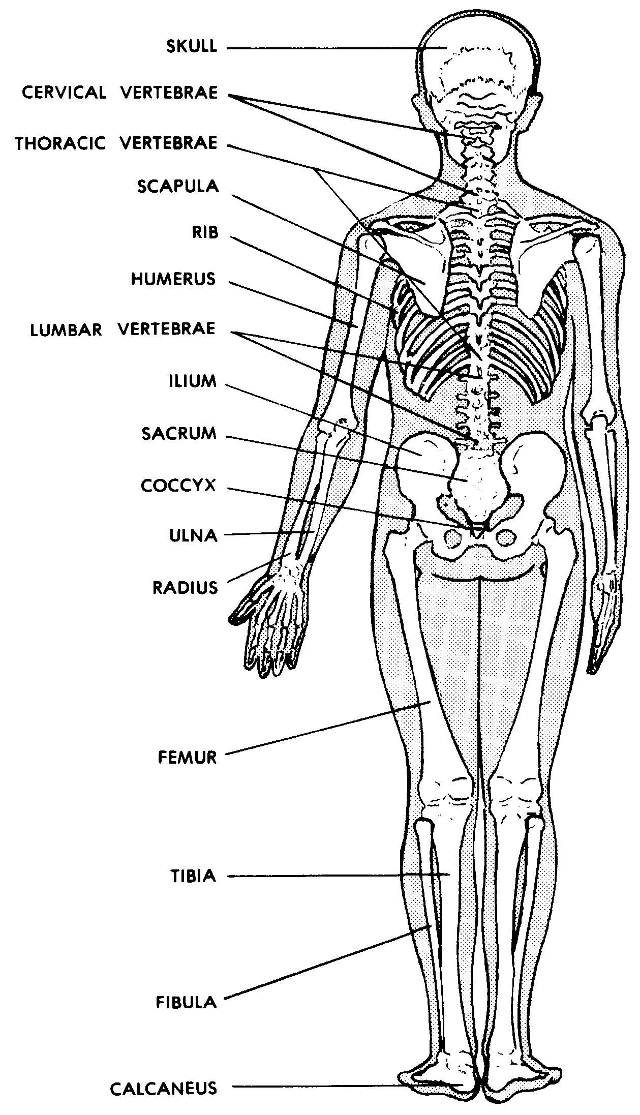
Images 04. Skeletal System Basic Human Anatomy
In addition, bones contain bone marrow and periosteum. You can see these tissues in Figure 4.4.2 4.4. 2. Bone marrow is a soft connective tissue that is found inside a cavity, called the marrow cavity. There are two types of marrow in adults, yellow bone marrow, which consists mostly of fat, and red bone marrow.
Divisions of the Skeletal System · Anatomy and Physiology
Key facts about the main bones, joints and muscles of the body. Axial skeleton: bones of the skull, ribs, vertebral column, sternum, sacrum, coccyx, hyoid bone and auditory ossicles. Appendicular skeleton: bones of the upper and lower limbs and the shoulder and pelvic girdles. Skull sutures, temporomandibular, shoulder, elbow, wrist, hip, knee.

Anatomy Of Long Bone Diagram A Typical Gross B On Human bones anatomy
These are (1) the axial, comprising the vertebral column —the spine—and much of the skull, and (2) the appendicular, to which the pelvic (hip) and pectoral (shoulder) girdles and the bones and cartilages of the limbs belong.
PreLab 2 Human Anatomy Lab Manual
Skeletal System: Labeled Diagram of Major Organs In addition to the bones, organs of the skeletal system include ligaments that attach bones to other bones and cartilage that provides padding between bones that form joints throughout your body.

Skeletal system 1 the anatomy and physiology of bones Nursing Times
The structure of a long bone allows for the best visualization of all of the parts of a bone ( [link] ). A long bone has two parts: the diaphysis and the epiphysis. The diaphysis is the tubular shaft that runs between the proximal and distal ends of the bone.

The Skull Anatomy and Physiology I
Anatomy of the Bone Bones and Joints What is bone? Bone is living tissue that makes up the body's skeleton. There are 3 types of bone tissue, including the following: Compact tissue. The harder, outer tissue of bones. Cancellous tissue. The sponge-like tissue inside bones. Subchondral tissue.
Long Bone Diagram Labled They are one of five types of bones
The structure of bones Tridsanu Thophet/EyeEm/Getty Images Bones are composed of two types of tissue. Compact (cortical) bone is a hard outer layer that is dense, strong, and durable. It.
File603 Anatomy of Long Bone.jpg Wikimedia Commons
Bones are your body's main form of structural support. They're made of hard, strong tissue that gives your body its shape and helps you move. Your bones are like the frame under the walls of your home. If you've ever watched a home improvement show and seen the internal structure of a house, that's what your bones are — the supports.

Skeletal system Quizizz
Sesamoid bones vary in number and placement from person to person but are typically found in tendons associated with the feet, hands, and knees. The patellae (singular = patella) are the only sesamoid bones found in common with every person. Table 6.1 reviews bone classifications with their associated features, functions, and examples.
