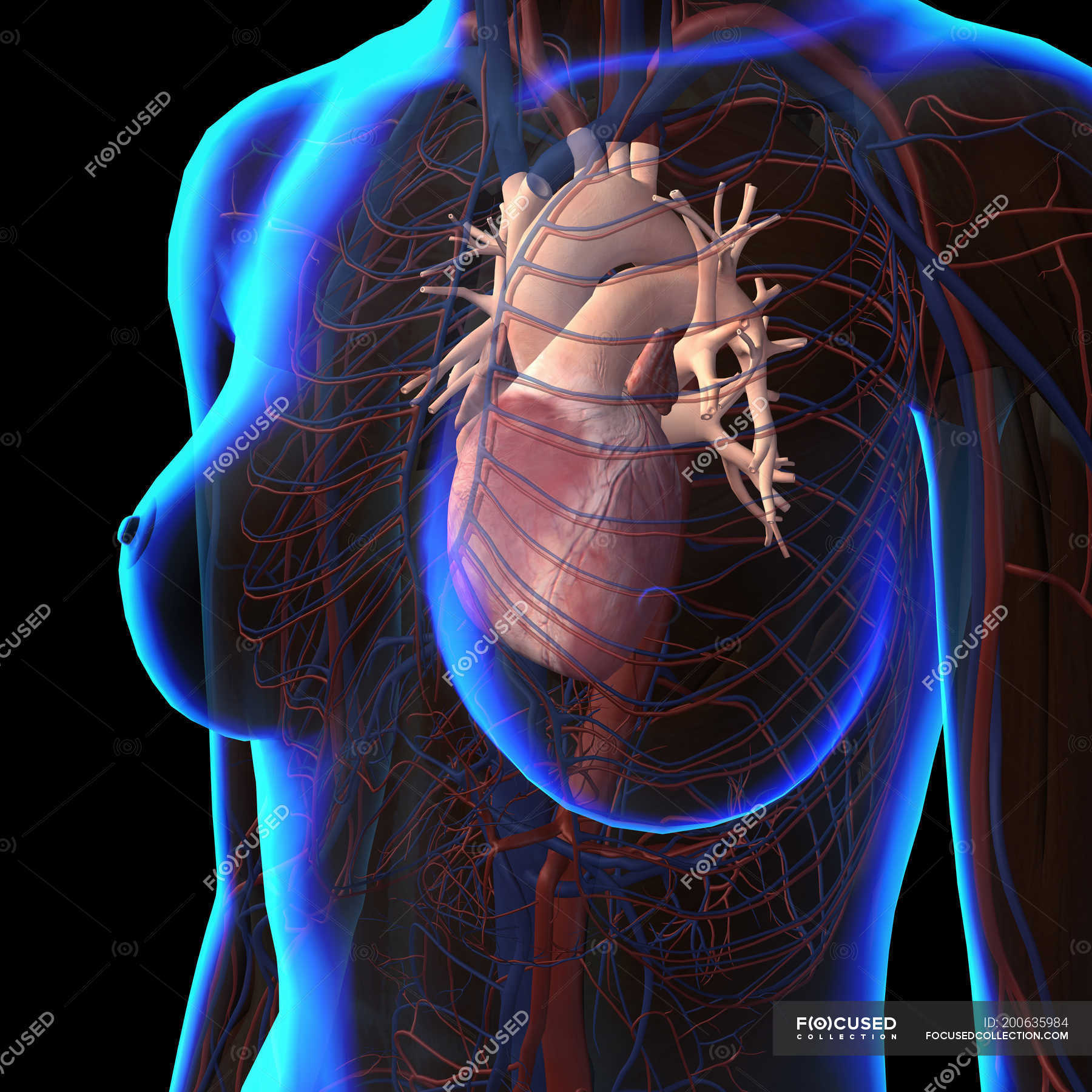
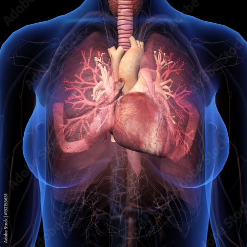
Xray view of female chest with heart and circulatory system — biology
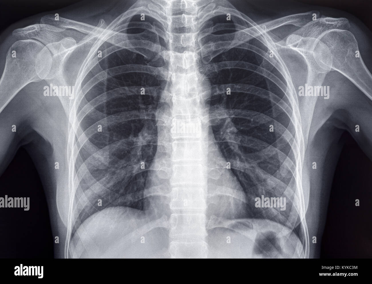
Lymph nodes, picture. A chest X-ray is a picture of the chest that shows your heart, lungs, airway, blood vessels, and lymph nodes. A chest X-ray also shows the bones of your spine and chest, including your breastbone, your ribs, your collarbone, and the upper part of your spine. A chest X-ray is the most common imaging test or X-ray used to.

Chest X Ray Female Front Stock Photos, Pictures & RoyaltyFree Images
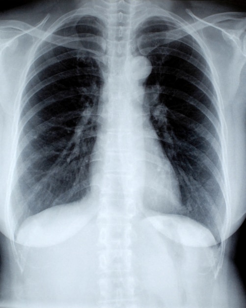
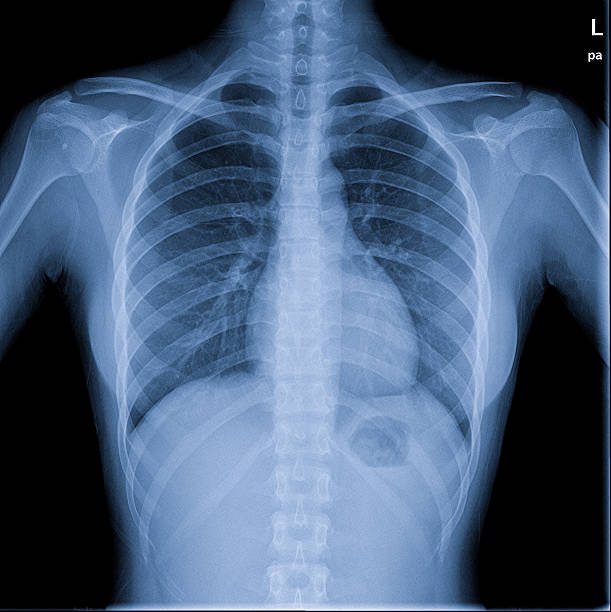
Chest X-rays produce images of your heart, lungs, blood vessels, airways, and the bones of your chest and spine. Chest X-rays can also reveal fluid in or around your lungs or air surrounding a lung.
Female chest x ray Photo Free Download
A chest X-ray helps detect problems with your heart and lungs. The chest X-ray on the left is normal. The image on the right shows a mass in the right lung. There is a problem with information submitted for this request. Review/update the information highlighted below and resubmit the form.

Female Chest X Ray submited images.
Benign and Malignant Tumours of the Female Reproductive System. 72. Normal Pregnancy. 73. Ectopic Pregnancy. 74. Placenta Previa. 75. Gynecology and Obstetrics - References. XII. Chapter 12 - Head and Neck.. Figure 9.1 Normal PA Chest x-ray, Labelled . Figure 9.2 Normal Lateral Chest x-ray, Labelled. ODIN Link for Normal Chest x-ray images,.
Chest Xray Of Female Photograph by Ted Kinsman Fine Art America
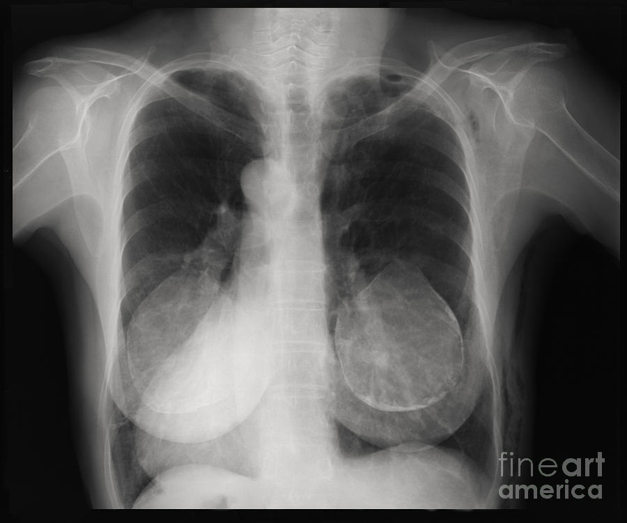
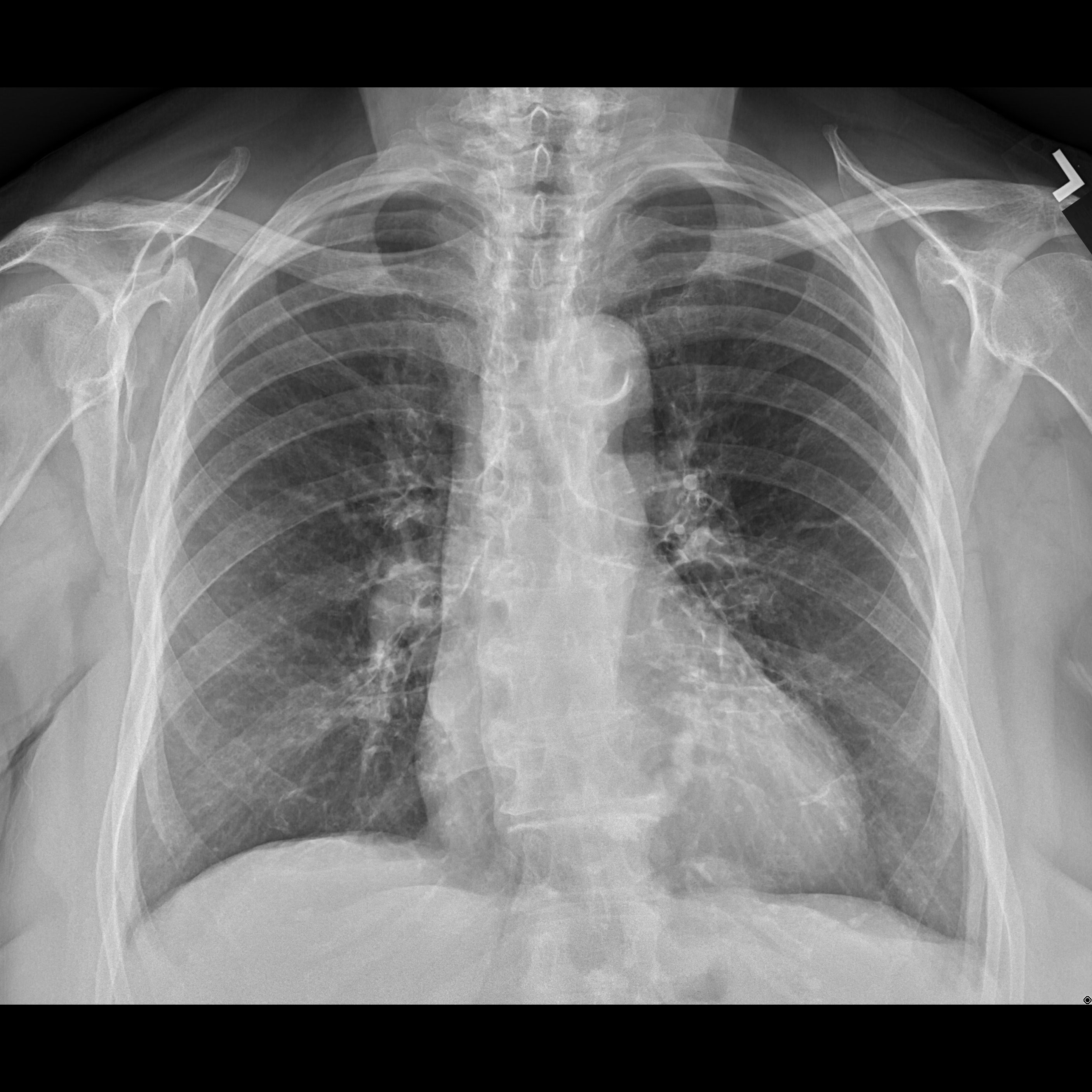
x-ray. This is a normal radiograph. The anatomy of the chest radiograph ( young adult female ) is well illustrated. Annotated image. Annotations of key anatomy: CPA = Costophrenic angle. BS = Breast shadow. SDS = Subdiaphragmatic space. CP = Cardiophrenic angle.
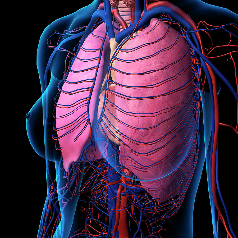
Xray View Of Female Chest, Heart Photograph by Hank Grebe Pixels
A chest x-ray produces images of the heart, lungs, airways, blood vessels and the bones of the spine and chest. An x-ray exam helps doctors diagnose and treat medical conditions. It exposes you to a small dose of ionizing radiation to produce pictures of the inside of the body. X-rays are the oldest and most often used form of medical imaging.

Chest Xray Female34 Image & Photo (Free Trial) Bigstock
Lung Cancer Screening. A chest X-ray creates images of: Lungs. Airways. Heart. Blood vessels. Bones of the chest and spine. It is often the first imaging test a doctor will order if lung or heart disease is suspected. If lung cancer is present, chest X-rays can sometimes detect larger tumors.

Young Woman Reveals Chest Xray Stock Photo Getty Images
A chest X-ray is an imaging test that uses X-rays to look at the structures and organs in your chest. It can help your healthcare provider see how well your lungs and heart are working. Certain heart problems can cause changes in your lungs. Certain diseases can cause changes in the structure of the heart or lungs.

Normal healthy Chest xray Stock Image C019/7286 Science Photo
A chest X-ray is a painless, noninvasive procedure with few risks. X-rays use a small amount of radiation, about the same levels that occur naturally in the environment. At Stanford, we take extra precautions to minimize our patients' exposure to radiation, including using: A protective lead apron to shield certain parts of the body.

Xray Film of Human Female Chest Stock Photo Image of radiology
A chest X-ray is a quick, noninvasive way for your provider to check the overall health of your lungs, heart and ribcage. It's often one of the first tests they use to diagnose broken bones or lung and heart conditions. And it can help your provider determine what kind of treatment you need. Medically Reviewed.
CXDI Digital Radiography Systems Clinical Gallery Canon Medical
A chest x-ray is a radiology test that involves exposing the chest briefly to radiation to produce an image of the chest and the internal organs of the chest. A normal chest x-ray can be used to define and interpret abnormalities of the lungs such as excessive fluid, pneumonia, bronchitis, asthma, cysts, and cancer.. Women who are pregnant.
"Female Chest Xray with Heart and Bronchial Tubes" Stock photo and
A chest X-ray shows the location, size and shape of the heart, lungs and some blood vessels.. This small amount of radiation isn't considered dangerous. However, pregnant women should avoid even this low level of radiation, when possible. Written by American Heart Association editorial staff and reviewed by science and medicine advisors.
Female Chest Stock Photos, Pictures & RoyaltyFree Images iStock
example 4 : adult male, arterial phase. example 5: adult female, arterial phase. HRCT chest. example 1 : non-contrast, supine, prone and expiratory. CT pulmonary angiogram (CTPA) example 1 : excellent pulmonary arterial / venous differentiation. example 2: average pulmonary arterial / venous differentiation. example 3: spectral CTPA.

The preoperative chest PA xray shows no abnormal findings in the lung
A chest X-ray test is a very common, non-invasive radiology test that produces an image of the chest and the internal organs. To produce a chest X-ray test, the chest is briefly exposed to radiation from an X-ray machine and an image is produced on a film or into a digital computer. Chest X-ray is also referred to as a chest radiograph, chest roentgenogram, or CXR.

Female Chest X Ray Royalty Free Stock Image Image 7011886
Fig. 3.7. Chest Versus Rib Technique. (A) A normal chest x-ray is taken at a relatively high voltage, allowing you to see the heart, pulmonary vessels, and skeletal structures. (B) By lowering the voltage of the x-ray beam, the pulmonary vessels become much harder to see and the bones become easier to see.
Chest Xray of an Adult Female Human Stock Photo Alamy
An X-ray is an imaging test that uses small amounts of radiation to produce pictures of the organs, tissues, and bones of the body. When focused on the chest, it can help spot abnormalities or.
