
Couinaud classification of hepatic segments Radiology Reference
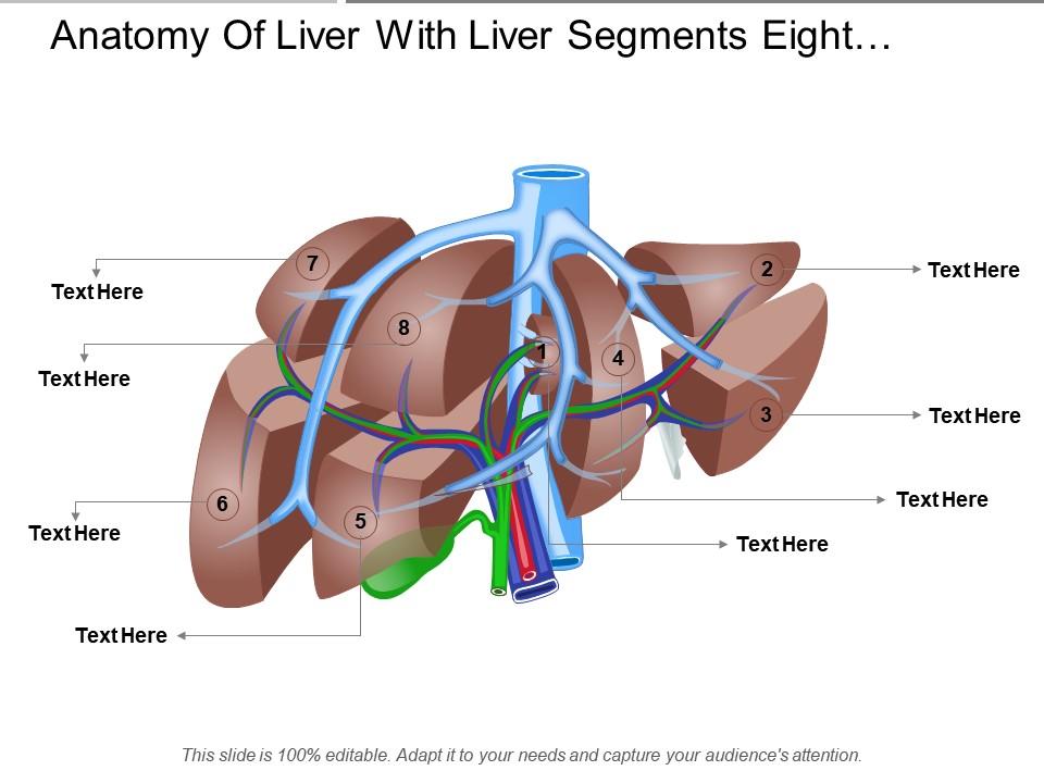
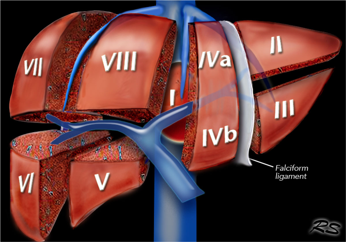
The most widely accepted model for liver segmental anatomy was first described by Couinaud (Couinaud 1957), and then popularised by Bismuth (Bismuth 1982; Bismuth et al. 1982; Majno et al. 2014), and is now the most commonly used in Europe. According to these authors, the liver is composed of eight anatomical segments, characterised by.

bismuth of liver Google Search Radiology, Ultrasound, Liver anatomy
Segments segment 1 (I) is the caudate lobe bounded posterolaterally by the fossa for the inferior vena cava, anteriorly by the ligamentum venosum, and inferiorly by the porta hepatis its inferior portion is subdivided into a lateral caudate process and a medial papillary process 6 may receive its supply from both the right and the left portal vein
Radiopaedia CTscan Hepatic segments coronal section labels
Hepatic segmentation Segmentatio hepatis Definition The hepatic segmentation (lobes, parts, divisions and segments) is the oganization of the liver into parts, divisions and segments.
Liver Radiology Key
Liver volume is assessed primarily via organ segmentation of computed tomography (CT) and magnetic resonance imaging (MRI) images. The goal of this paper is to provide an accessible overview of liver segmentation targeted at radiologists and other healthcare professionals. Methods

Couinaud’s Liver Segments Sonographic Tendencies Segmentation
Segmental anatomy Traditionally, the liver was divided into four anatomical lobes: the right, left, caudate and quadrate lobes.

Segmental Anatomy Of Liver
Liver Segments (Axial & Coronal) by R. Furman Borst MD; Abdo by Whitney Graff; Anatomía by Diego González; Normals by Noah; ASA 2017 Abdominal anatomy refresher by Craig Hacking Gen Surg by Aaron Ow; Liver Prokop by Stefan Teodoru-Saman; ASA Radiopaedia for sonographers by Craig Hacking Anatomy by Vitalii Rogalskyi; Anatomy by muhammet
Liver Anatomy Segments Anatomical Charts & Posters
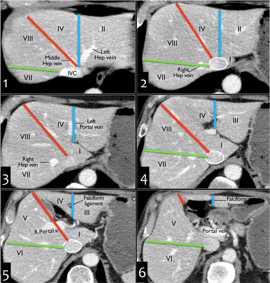
Segmental anatomy according to Couinaud. Use your right fist to represent the liver segmental anatomy. Para-sagittal Left Ultrasound image. Sagittal Midline Ultrasound image. The Ligamentum venosum is highlighted in orange. lhv:Left hepatic vein ivc: Inferior vena cava Ultrasound image- Para-sagittal Midclavicular.

Couinaud classification of hepatic segments Radiology Reference
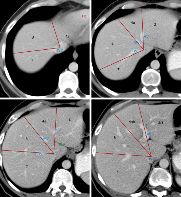
In a portal venous phase CT, the right, middle, and left hepatic veins are usually filled with contrast-mixed blood and can be seen emptying into the inferior vena cava, which is found along the back side of the liver. The portal and hepatic veins divide the liver into segmental anatomy that allows radiologists and surgeons to be very specific.
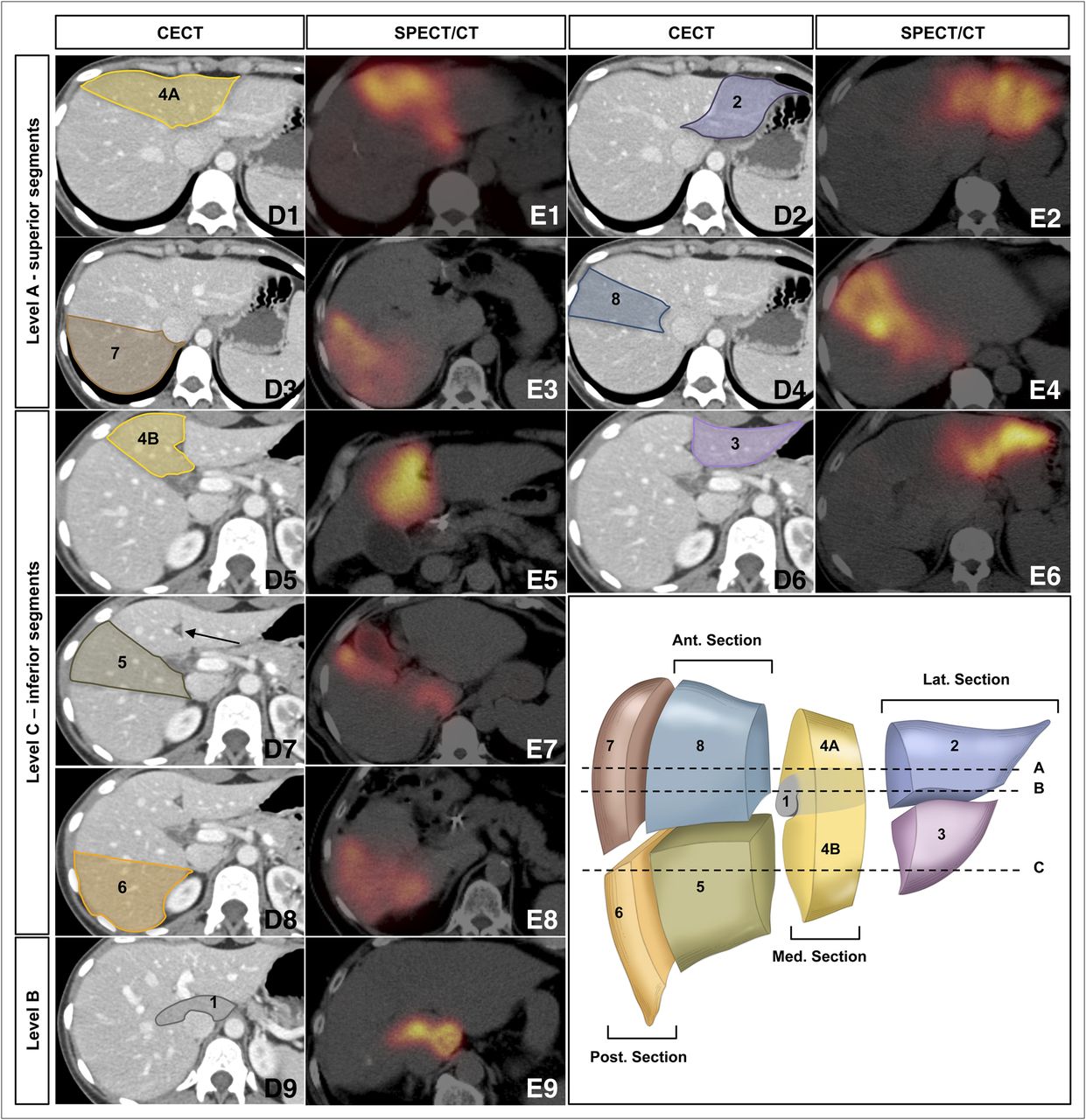
From the Angio Suite to the γCamera Vascular Mapping and 99mTcMAA
The authors developed a simplified description of the segmental anatomy of the liver on the basis of the Couinaud nomenclature. This approach was demonstrated with normal in vivo sonographic images and livers dissected in corresponding planes. The branches of the portal vein, which lead to the center of individual segments, are used as key landmarks in the determination of segmental location.

Couinaud classification of hepatic segments Radiology Reference
Indications Liver ultrasound is commonly utilized in the evaluation of various hepatic conditions. The most frequent indications include 1,2: screening for hepatic disease in at-risk populations (e.g. hepatocellular cancer screening in cirrhotic patients) investigation of abnormal liver function tests

Couinaud’s Liver Segments Sonographic Tendencies Segmentation
1 case No description provided Article Couinaud classification of hepatic segments The Couinaud classification (French eponym: pronounced kwee-NO) is currently the most widely used system to describe functional liver anatomy.
Liver Anatomy Segments
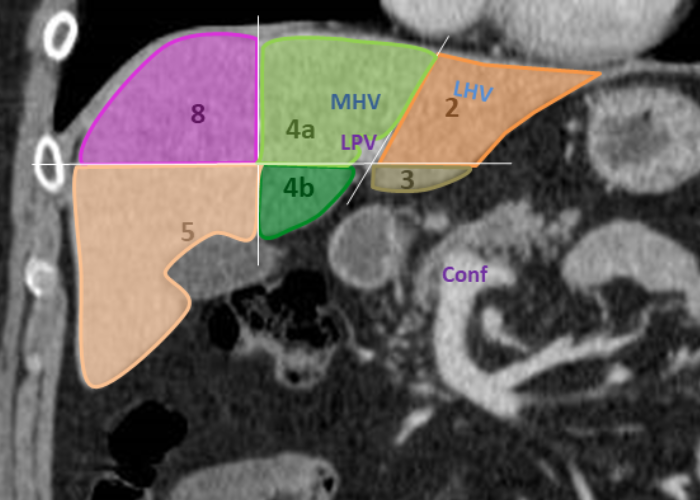
Segmental anatomy according to Couinaud. Click to enlarge. Couinaud classification The Couinaud classification of liver anatomy divides the liver into eight functionally indepedent segments. Each segment has its own vascular inflow, outflow and biliary drainage.

Couinaud classification of hepatic segments Radiology Reference
The liver has the capacity to store enough vitamin A to prevent a deficiency for as long as 1-2 years 4 and enough vitamin D and vitamin B 12 to prevent deficiency for 1-4 months. The liver also stores glycogen, fats, and amino acids and can metabolize them into glucose or vice versa, depending on the body's needs.

Liver Anatomy Segments
The location of each hepatic segment can be estimated if the artery supplying the segment can be correctly identified on angiograms. Notably, morphologic differences in the hepatic artery system caused by respiration should be recognized. Supplemental material available at http://radiographics.rsna.org/lookup/suppl/doi:10.1148/rg.e37/-/DC1

Radiology Assistant Liver Segments
A liver segment is one of eight segments of the liver as described in the widely used Couinaud classification (named after Claude Couinaud) in the anatomy of the liver.This system divides the lobes of the liver into eight segments based on a transverse plane through the bifurcation of the main portal vein, arranged in a clockwise manner starting from the caudate lobe.
Сегменты печени схема анатомия
Abstract The availability of intra-arterial hepatic therapies (radio and/or chemo-embolisation, intra-arterial hepatic chemotherapy) has convinced radiologists to perfect their knowledge of the anatomy of the liver arteries. These sometimes, complex procedures most often require selective arterial catheterization.
